A dental implant treatment is not a single act but a sequence of clinical decisions and coordinated steps that begin long before surgery and continue long after the crown is placed. Thinking of it as a process rather than a product helps patients and clinicians align expectations, plan for contingencies, and focus on outcomes that last. The end goal is straightforward: a tooth that looks natural, functions comfortably, and stays healthy in the mouth for many years. Achieving that goal requires careful assessment, transparent planning, meticulous execution, and committed maintenance.
Every complete implant journey starts with diagnosis. This means a thorough medical and dental history, discussion of goals, and a clinical examination of teeth, gums, bite and jaw function. The clinician assesses gum health, measures pockets, checks for mobility, notes any caries, existing restorations and parafunctional habits such as clenching or grinding. The medical history is equally important. Uncontrolled diabetes, smoking, certain medications, autoimmune conditions, prior head and neck radiotherapy, and bleeding disorders can alter healing and risk profiles. None of these automatically disqualify a patient, but they shape the plan. Setting a baseline at this stage also allows later comparisons and serves as an informed consent foundation.
Imaging is the next pillar. A panoramic radiograph offers a broad overview of both jaws, sinuses, and critical anatomical landmarks. For implant planning, a cone beam computed tomography scan, often called a CBCT, is the modern standard. The CBCT provides three dimensional information about bone width, height and density, the location of the mandibular canal and mental foramen, and the shape of the maxillary sinus. It also reveals hidden pathology such as cysts or impacted roots. With this information, the clinician can determine whether bone augmentation is necessary, select implant dimensions, and anticipate the ideal angulation. When available, digital impressions of the teeth and gums are combined with the CBCT to simulate the restoration in the correct position first, then fit the implant into that plan. This restorative driven planning avoids the common mistake of placing implants where bone is easiest rather than where the prosthetic tooth needs to be.
Treatment planning is the stage where patient priorities and clinical realities are reconciled. The plan should explain the number and position of implants, whether extraction and site preservation are needed, whether bone grafting or sinus elevation is recommended, and the timeline from surgery to final crown. It should identify the type of restoration, for example a single crown, a multi unit bridge, or a full arch fixed prosthesis, and indicate whether the provisional phase will be fixed or removable. The plan sets expectations about aesthetics, especially in the front of the mouth where gum contours, smile line and symmetry demand precision. It addresses occlusion and jaw function, since implants do not have the ligamentous cushioning of natural teeth and are less forgiving of overload. A good plan also defines maintenance responsibilities and follow up intervals. Cost transparency is part of a complete plan. Itemizing imaging, surgery, grafting, components, provisional, final restoration and maintenance avoids surprises.
The consent conversation turns that plan into a shared commitment. It should cover the benefits and alternatives such as a traditional bridge or removable partial denture, along with risks like infection, nerve injury, sinus complications, lack of osseointegration, aesthetic compromise and long term peri implant disease. Patients should be told how long the different phases take, what the temporary tooth will be like, whether any time without a tooth is expected, and what the rules will be for diet and hygiene during healing. Consent is most useful when it is paired with clear instructions that the patient can take home.
When a failing tooth is present, extraction strategy is central. Sometimes a tooth can be removed atraumatically with immediate implant placement into the same socket. This saves time and helps preserve bone and soft tissue contours. Immediate placement requires strict criteria: the socket must be infection free or well managed, sufficient bone must be present for primary stability, and the implant angulation must allow a future restoration that is cleansable and harmonious with the bite. If these criteria are not met, a staged approach is safer. This might include socket preservation with a bone substitute and membrane to maintain ridge volume, a healing period of several months, then implant placement once the site is stable. Patients often prefer faster schedules, but the anatomy and risk profile make the decision.
Bone augmentation is a topic that deserves a simple explanation. Implants need bone around them to integrate and to be stable under chewing forces. If your bone is too thin or too short at the planned site, augmentation can rebuild the foundation. This might be a minor contour graft at the time of implant placement, or a separate procedure such as a lateral sinus floor elevation in the upper posterior jaw or a block graft in the lower jaw. Materials range from autogenous bone to processed grafts and synthetic substitutes, with resorbable or non resorbable membranes to guide healing. Augmentation extends the timeline but often converts a compromised site into a predictable one.
The day of surgery begins with aseptic protocol and pain control. Options include local anesthesia, oral sedation, intravenous sedation, or general anesthesia depending on complexity and patient preference. A full sterile field is not always necessary in office based implantology, but the principles of clean instruments, draping, and a controlled environment reduce contamination. When guided surgery is used, a custom surgical guide translates the digital plan to the mouth and helps place the implant at the intended angle and depth. Freehand surgery remains common and effective in skilled hands. In both approaches, the bone is prepared with calibrated drills or piezoelectric instruments according to the implant system’s sequence, and the implant is inserted to a torque and stability threshold chosen by the clinician.
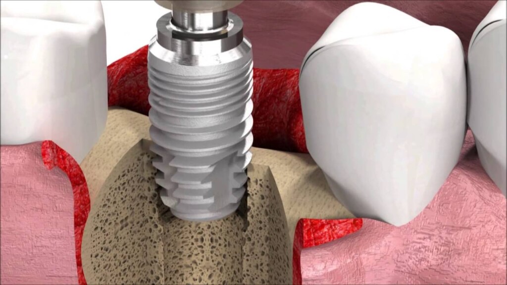
Implant components can be demystified. The fixture is the threaded part that resides in the bone and becomes the artificial root. The abutment is the connector that emerges from the gum and provides a platform for the crown. A screw joins the abutment and the implant or secures the crown directly to the implant in screw retained designs. There are healing abutments to shape the gum during integration, temporary abutments to support provisional crowns, and final abutments customized for proper emergence profile. The connection geometry matters because it determines component compatibility, screw mechanics and load distribution. Patients do not need to memorize brand catalogues, but they should expect traceability of the implant system, records of part numbers and lot numbers, and assurance that future maintenance will be possible.
Immediate temporization is sometimes offered, meaning a temporary crown is placed the same day as the implant. This is common in the aesthetic zone and in full arch protocols when stability is excellent and the bite can be controlled. The temporary is designed out of occlusion or with carefully planned contacts to protect the implant while it integrates. Other cases use a small healing abutment with no tooth on it and a removable provisional to fill the gap during healing. Either method can be correct; the criterion is biologic safety. Patients should be informed that immediate teeth are for appearance and comfort, not for heavy chewing at first.
The healing phase, called osseointegration, is when bone cells grow onto the surface of the implant. This typically takes several weeks to a few months depending on bone quality, patient factors and whether grafting was performed. During this time, the soft tissues around the implant are shaped to create a natural emergence profile. In the front of the mouth, the provisional is often adjusted several times to refine gum contours in a gentle, progressive way. Pictures and shade records may be taken so the final crown can be matched to the surrounding teeth.
The prosthetic phase converts the integrated implant into a functional tooth. It begins with an impression or a digital scan. Conventional impressions use impression copings attached to the implant and elastomeric materials to capture their position. Digital impressions use scan bodies and an intraoral scanner to map the 3D relationship without trays or putty. Both methods aim to record the implant position, the gum shape, the bite and the appearance of adjacent teeth. The laboratory then designs the abutment and crown. Abutments can be stock, meaning prefabricated in standard shapes, or custom milled from titanium or zirconia to match the patient’s gum contour and the planned crown geometry. Crowns can be monolithic zirconia for strength, layered porcelain for subtle aesthetics, or high strength composite in specific indications. In the aesthetic zone, a custom abutment with a layered ceramic crown often delivers the most lifelike result.
When choosing between screw retained and cement retained crowns, maintenance and risk drive the decision. Screw retained crowns allow easy removal for hygiene or repair and avoid residual cement in the sulcus, which can inflame the tissues. Cement retained crowns can help in situations with challenging angulation, but they demand controlled cementation protocols and often the use of vent holes or retrievability strategies. In full arch cases, the most common modern solution is a screw retained bridge on four to six implants per jaw, carefully designed to be cleansable and to distribute forces evenly. The clinician will check fit and passivity, confirming that the framework seats without inducing stress.
Occlusion, the way teeth contact and move, is a silent determinant of long term success. Because implants lack the shock absorbing ligament of natural teeth, they should not be the first point of contact in a heavy bite, and excursive movements should not overload them. In single tooth cases, the crown is adjusted to share load appropriately with neighbors. In full arch cases, the occlusal scheme is designed to stabilize jaws while protecting implants and opposing structures. For patients with bruxism or clenching, a night guard is often prescribed once healing is complete. This simple device can dramatically reduce fracture and screw loosening.
Hygiene training turns a finished reconstruction into a durable one. Patients should be taught how to brush around the implant neck and under any pontic areas, how to use interdental brushes, floss or specialized threaders, and how to clean under a full arch bridge where food can collect. The goal is to keep the mucosa healthy and the biofilm controlled. Early signs of inflammation such as bleeding on probing or persistent redness should trigger professional intervention. A recall schedule might begin with reviews at two weeks for soft tissue checks, at two to three months for occlusion and hygiene review, and then every four to six months for maintenance depending on risk.
Documentation is an often overlooked part of a complete treatment. Patients should receive a summary of the plan and the actual procedures performed, a list of implant system, sizes, abutments and torque values used, radiographs showing the implant and crown in place, and instructions for maintenance. This record facilitates care in the future if the patient moves or another clinician needs to troubleshoot a problem. In cross border care, documentation is essential because compatibility of parts and communication between teams protects the investment.
Complications can occur even in well planned cases. Early problems include postoperative pain beyond expected levels, bleeding, infection at the incision, or stability concerns. Later problems include screw loosening, crown fracture, ceramic chipping, gum recession, and peri implant mucositis or peri implantitis, which is inflammation with or without bone loss. A complete treatment plan includes an approach for these issues. For example, a screw retained crown that becomes loose can be tightened with a new screw at the proper torque after checking occlusion. A chipped veneer can sometimes be polished or repaired. Inflammation is managed with debridement, improved hygiene, and, if needed, adjunctive antimicrobials or surgical therapy. The best strategy remains prevention by designing cleansable restorations and maintaining regular professional care.
Materials deserve a brief discussion. The implant fixture is almost always titanium alloy with surface treatments that accelerate bone integration. Zirconia implants exist and can be useful for specific allergies or aesthetic preferences, but they require careful case selection. Abutments may be titanium for strength, titanium with ceramic sleeve for aesthetics, or monolithic zirconia in selected positions. Crowns can be high strength monolithic zirconia for posterior durability, layered porcelain fused to zirconia for anterior translucency, or nano ceramic composites for shock absorption in particular cases. The right choice balances aesthetics, space, wear on opposing teeth, and risk of fracture. The patient’s habits guide whether toughness or visual finesse should be prioritized.
Timelines are best expressed as ranges because biology varies. In straightforward single tooth cases with adequate bone, the path from implant placement to final crown might be eight to twelve weeks, extended if grafting was simultaneous. In immediate placement with immediate provisional, the patient leaves with a temporary but still waits for integration before receiving the final crown. In graft heavy or sinus lift cases, the journey might take six to nine months. Full arch immediate load protocols deliver a fixed provisional within days, followed by a definitive bridge after three to six months once tissues stabilize. Setting these expectations early reduces frustration and improves satisfaction.
Pain control and comfort are integral to a complete experience. Beyond anesthesia during surgery, patients should be counseled on what to expect afterward, how to use analgesics, when to apply cold packs, what to eat, and how to sleep to reduce swelling. They should be given a hotline or clear instructions for concerns such as increasing pain, fever, or unusual bleeding. Most implant patients are pleasantly surprised by how manageable discomfort is when the procedure is well planned and carried out with minimal trauma.
Financial planning is part of completeness. Implants are an investment and costs can escalate if ancillary procedures are needed. A comprehensive quote should separate diagnostics, surgery, grafts, components, temporaries, final prosthetics, and maintenance. Policies for warranties or remakes should be spelled out. Some clinics offer staged payments that align with treatment milestones. Understanding the financial structure aligns interests and prevents the temptation to cut clinical corners to fit a number.
Choosing the team matters as much as choosing the implant. Ideally, surgical and prosthetic phases are coordinated, whether by the same clinician or by a close collaboration between a surgeon and a restorative dentist. A skilled laboratory is an indispensable partner because the abutment design, crown morphology and shade layering translate clinical intent into reality. The best outcomes happen when communication flows both ways, with photos, shade tabs, digital designs and even try ins used to refine the result. Patients should feel comfortable asking who does what and how the team collaborates.
The final delivery of the crown is the culmination but not the end. Fit is verified clinically and radiographically. Contacts with neighboring teeth are checked so food does not trap but floss can still pass. The bite is adjusted in static closure and in movements. The patient is taught how to clean, what to expect in the first week, and when to return. A follow up a few weeks later catches any minor issues after the mouth has adapted. The long term plan includes periodic professional cleanings with instruments and techniques safe for implants, tissue and bone level monitoring, and updates to the night guard if wear is observed.
It is helpful to summarize the essential attributes of a complete implant treatment. It begins with a patient centered diagnosis and a CBCT based plan that is restorative driven. It explains alternatives and risks transparently and gains informed consent. It preserves or rebuilds bone and soft tissue as needed rather than forcing a quick fix. It uses components that are traceable and compatible long term. It manages the provisional phase to sculpt healthy, natural contours. It delivers a prosthesis that fits passively, functions comfortably, and can be maintained. It teaches the patient how to care for the restoration and invites the patient back regularly for professional maintenance. It documents every significant decision and step so future care is seamless.
When all of these elements are present, the implant does what patients hope it will do. It disappears into the smile, it lets them eat without thinking, and it remains stable in health. This is the quiet success of modern implant dentistry. It is not magic and it is not luck. It is the product of careful planning, excellent execution and shared responsibility between the dental team and the patient. From the first radiograph to the last polish on the final crown, completeness is what turns an implant from a metal fixture into a reliable, natural part of everyday life.